Skip to content

Dr Randy Kyes Retires After 35 Years of Global Conservation, Education and Empathy

Dr. Kyes and wife Elle in Thailand in 2021.

After 35 years with the Washington National Primate Research Center (WaNPRC) and the University of Washington, Dr. Randall (‘Randy’) Kyes, Unit Chief of the Center’s Unit of Global Programs, Research Professor in the Dept. of Psychology, adjunct in the Depts. of Global Health and Anthropology, and Director of the Center for Global Field Study, will retire at the end of December 2025. His career leaves behind a legacy of science, service, and compassion that spans continents and generations.

From the tropical forests of Indonesia to the mountains of Nepal, Kyes has devoted his entire professional life to building lasting international partnerships and training thousands of students and professionals to care for the natural world and one another.

“Everything we do to the environment affects our own health,” he said. “If we can improve environmental health, we improve human health too.” That belief was the foundation for decades of work connecting people and ecosystems across the globe.

Dr. Kyes (left) and local staff during his first year on Tinjil Island (1990) conducting a population survey of the macaques.

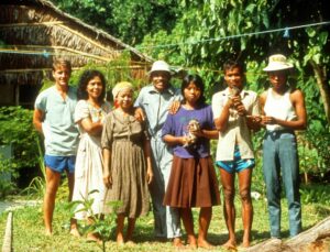
When asked to look back, Dr Kyes seems amazed at how it all began. In 1989, during his postdoc at Wake Forest School of Medicine, he met an Indonesian scientist, Dondin Sajuthi, who mentioned a small island where long-tailed macaques were being released to establish a breeding population. “He told me the folks at the University of Washington were looking for someone to monitor how the monkeys were doing,” Kyes recalled. “I went the next summer to conduct a population survey of the macaques, and that was it. Everything started from there.”

That was Tinjil Island, Indonesia. In 1991, Dr Kyes led his first field course there, an experience that would grow into a global model for collaborative field training. Over the next three decades, he and his longtime collaborators, both international and domestic, led 148 field courses in eight countries for nearly 2,900 participants representing 20 nations.

“Our goal was always to help local people build their own capacity to manage their environment and health,” he said. “These programs were never about us showing up to teach and then leaving.  They were about partnership and long-term collaboration.”

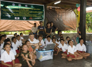
That philosophy extended well beyond university students. Around 2000, while working in a rural Indonesian village coping with crop-raiding monkeys, Dr. Kyes and his team met with village leaders to discuss ways to help mitigate the conflict and protect the endangered animals. “They were grateful, but they said, ‘We’re set in our ways. You should talk to our kids before they are, to help them appreciate the need to protect these monkeys?’”

He and his colleagues took that advice to heart. The following year they launched their first K-12 outreach program, combining lessons on wildlife conservation with art and health activities. Over the next 25 years, that grew into 181 outreach programs reaching more than 10,000 children across eight countries.

“One of my favorite moments came years later,” he said. “Two young women from that same village joined one of our field courses and told us they’d been third graders in our very first outreach session. Seeing that full circle, kids who grew up to become conservation students – that’s the kind of impact you hope for.”

Asked what kept him going through the endless travel, the weather, and the occasional political unrest, and he’ll point to his students. “Watching them discover the field, that spark when they realize they can do this work, is what I’ll miss the most,” he said.

The field courses, which combine research, cross-cultural exchange, and a dose of physical endurance, are intentionally small, he said. “We’re often in remote, challenging places. You learn to improvise, listen, and collaborate,” he said. “It’s not just about training and data collection.  It’s about experiencing the world first hand, while appreciating the similarities across all cultures.”

Running global programs for decades meant confronting real-world challenges. Some years were canceled due to political unrest or natural disasters. In 2015, an earthquake and landslide wiped out an entire Nepali village where Dr. Kyes and his colleagues had worked for more than a decade. “We knew those families,” he said quietly. “It was heartbreaking. But we went back. You don’t just walk away from people who’ve become part of your life.”

Those long-term connections remain what he values most. “I’ve stayed in people’s homes, watched their kids grow up,” he said. “That’s the reward, those friendships that cross borders.”

Incoming Associate Director Matthew Novak with Randy and Entang Iskandar, a student from Randy’s first field course, who has collaborated on all the field courses and who plans to continue that work.

He’s also quick to credit his wife and colleague Dr. Pensri (‘Elle’) Kyes, who has been part of the collaborative team since 2010 and became a Research Scientist with the Primate Center in 2014. She has been integral in helping run education programs and coordinate field logistics. “The outreach activities in many of our program countries wouldn’t exist without her,” he said. “She has an incredible way of connecting with communities and the children.”

Kyes’ work has always focused on empowerment. Every field course is designed and led with local partners who provide lectures, field expertise, and cultural context. That also helped create a network of alumni, many of whom now run their own training programs.

“I’ve been incredibly fortunate,” he said. “Few people get to do work that means so much to them personally and that connects them to so many others. It’s been the privilege of my life.”

Even as he retires from his formal UW roles, he hopes the programs will continue. “What I’d really love,” he said, “is to see the next generation keep this momentum going. If we’ve done our jobs right, they already are.”

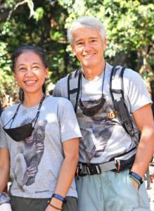
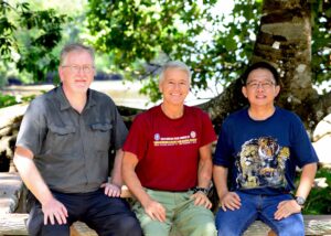
  • Randy and Elle pause during a field course, 2021

By the Numbers (1990–2025):

  • 35 years with the WaNPRC
  • 148 field courses in 8 countries
  • 2,894 participants representing 20 nations
  • 181 outreach programs for 10,272 K–12 students
  • 30+ years of continuous collaboration in Indonesia
  • 20+ years of continuous collaboration in Nepal and Thailand
  • 10+ years of collaboration in Bangladesh, China, India and Mexico

Related Links:

Neuro Unit Shines at SfN

SfN logo with an image of a brainResearchers from the Washington National Primate Research Center Neuroscience Unit made a big impact at this year’s Society for Neuroscience (SfN) meeting, sharing 27 different scientific presentations. This large wide-ranging set of contributions shows how our teams are helping push neuroscience forward in meaningful, practical ways.

The studies covered everything from basic brain wiring to vision, learning, and new tools that can change how scientists study the brain. Here are some of the standout projects:

Why different parts of the brain learn differently
One project showed that two neighboring brain areas, one that helps control movement and one that processes touch, don’t adapt to stimulation in the same way. This suggests that future brain-based therapies, like those used after injuries or strokes, may need to be tailored to each specific region rather than using a one-size-fits-all approach.

How the brain keeps the world “steady” when your eyes jump around
Every time you move your eyes, your visual world should technically blur. But it doesn’t; your brain keeps things stable. WaNPRC scientists uncovered signals in a part of the brain involved in vision that help update this “steady view” even when the eyes move rapidly. Understanding this process can eventually help with conditions that affect visual stability.

How the brain finds objects in cluttered scenes
Researchers tested how monkeys identify animals in very challenging pictures, like those made of dots or high-contrast black-and-white shapes. They found that mid-level visual areas and the prefrontal cortex work together to pull meaningful objects out of visual noise. This offers clues about how we make sense of complicated images in the real world.

How the brain recognizes shapes even when lighting changes
Another team discovered that a key visual area continues to recognize an object’s shape even when its brightness flips from light-on-dark to dark-on-light. This means the brain stores shape information in a way that’s tied to the object itself, not the lighting—similar to how you can recognize a friend’s face whether they’re in sunlight or shadow.

What changes across layers of the brain’s “first stop” for vision
Using recordings from thousands of neurons, researchers found that the way cells respond to overlapping patterns (like stripes or textures) changes depending on their depth within the visual cortex. This gives scientists a clearer map of how early visual processing works.

A breakthrough tool for turning brain activity off with light
One exciting advance this year was a new optogenetics technique that uses red light to temporarily quiet specific brain cells—no surgery required beyond the original gene delivery. The effect lasts more than a year and works through the brain’s protective covering. This is a major step forward for long-term, minimally invasive brain research.

The first direct look at how the brain filters visual information during eye movements
Scientists identified individual pairs of connected cells between the eye and the first visual relay in the brain. They found that this connection briefly “dials down” signals right before an eye movement, then opens back up afterward—almost like a camera adjusting exposure during motion. This helps explain how the brain avoids blurry vision.

Big steps toward mapping the brain’s wiring in multiple species
Two presentations showed how barcoding technologies  – tools that label neurons with unique molecular “IDs”-are being adapted for use in monkeys and other species. These methods could make it possible to map brain wiring at a massive scale.

How the brain gets better at learning new tasks
In a long-term study, researchers found that as animals learned many different problems over time, brain cells in memory and decision-making areas started representing information in more flexible, general ways. This work helps explain how we develop “learning-to-learn” skills, being able to pick up new tasks more easily with experience.

Together, these presentations highlight just how much groundbreaking work WaNPRC researchers are contributing to the field. Their discoveries deepen our understanding of how the brain sees, learns, adapts, and connects, and they pave the way for future advances in treating human neurological conditions.

This year’s SfN meeting confirmed what we already knew: our scientists are helping shape the future of neuroscience.

Proposed Federal Cuts to Animal Testing Spur Scientific Debate

green background with "STAT" lettered across the front.The NIH and FDA efforts to shift away from animal testing, promoting “new approach methodologies” (NAMs) like AI, organoids, and organ-on-a-chip systems are not finding unanimous support in the scientific community. The goal is to improve research efficiency, lower costs, and reduce harm to animals. While some scientists support the move as overdue, others warn that NAMs can’t yet replace animal models in many areas, like cancer and radiation research. Experts are also concerned about the speed of implementation and the risk of compromising scientific rigor. The shift comes amid broader federal cuts to basic science funding, raising doubts about how far these changes can be effectively realized.

You can read the entire article in STAT News here.

“The reason we have made all the progress and have such amazing science that we do today is in large part thanks to the animal models,” Sally Thompson-Iritani, Assistant Vice Provost of animal care at the University of Washington, told STAT.

“The NIH decided to put this name on it, ‘new approach methodologies,’ but they’re not new. We’ve been doing this for decades, constantly working in parallel,” Deborah Fuller, Director of the Washington National Primate Research Center at the University of Washington, told STAT.

Evidence of Bipartisan Support for Research in D.C.

screenshot of the livestream of the senate hearing. it depicts a dais for senators at the back of the image and a hearing room of seats before themI recently returned from Washington, D.C., where I was pleased to see something uncommon right now: bipartisan support for biomedical research. I want to applaud it and call out what WaNPRC is doing to continue to deserve that support.

I heard Republicans and Democrats alike praise the benefits of research at a Senate Appropriations Committee hearing entitled: Biomedical Research: Keeping America’s Edge in Innovation. Both the chair and vice chair, Sen. Susan Collings (R) Maine and Sen Patty Murray (D) Washington, spoke forcefully on the need for the federal government to continue funding biomedical research.

Senator Collins pointed out that every dollar of funding from the National Institutes of Health generates new economic activity of more than $2.50.  But the imperative to keep investing is “not just about scientists and researchers and economic activity,” she said.

“If clinical trials are halted, research is stopped. And laboratories are closed. Effective treatments and cures for diseases like Alzheimer’s, Type 1 Diabetes, childhood cancers, and Duchene’s Muscular dystrophy will be delayed or not discovered at all. We must preserve and strengthen America’s leadership for the sake of families all across this country,” she said.

Senator Murray concurred, pointing to the obvious benefits of government-backed research.  “Those investments have paid off in so many ways. Not just billions in economic activity, hundreds of thousands of jobs and a medical research enterprise that is the envy of the world. They’ve also paid off with genuine miracles. Cures that were once impossible, treatments that were once unthinkable. These are investments that give patients hope for the future, that give them back a live derailed by a disease, that give people precious more time with their loved ones. Which is why I am so deeply alarmed that President Trump has taken a wrecking ball to our biomedical research enterprise.”

Senator Murray was referring to attempts to gut funding by cutting indirect costs that provide the very buildings and equipment and other support that researchers need to do this work, and by attempts to slash contracts and gut agencies that fund research and protect public health.

As I sat in the chamber, I was deeply moved by the personal story of one witness at the hearing. Emily Stenson is a mom whose daughter Charlie was three when she was diagnosed with cancer. With Charlie, now five, on her lap, Ms. Stenson testified that her daughter, whose cancer is in remission, was benefiting from a new mRNA-backed test to detect relapse.  A test discovered through research at Seattle Children’s Hospital and the University of Washington.

“We first learned about this test in 2023, and knowing it’s almost within reach brings us so much hope. It’s real proof that continued research doesn’t just change lives—it saves them,” she said. Then she added, “…if you were in my shoes, you would want to know that everything possible was being done to save your child or grandchild…. Cuts to medical research are not just numbers on a spreadsheet—they are stolen chances, unfinished stories, and futures left unrealized… That is why I am asking you today: please continue to fund and prioritize pediatric medical research. Every investment you make saves lives, protects futures, and gives children like Charlie a fighting chance to grow up and thrive.”

At WaNPRC, we are deeply motivated by the impact our research has on human and animal health. Some of us are motivated by personal experience, having benefited from that research.

And while the administration vows to phase out animal testing requirements for certain treatments, I believe we can be part of a future that contributes to reaching the goal of using New Approach Methodologies (NAMs) that can reduce animal  testing. Right now, it’s not clear if NAMs can ever fully replace animal testing, but it’s becoming clearer that it can help reduce it. Examples include using Artificial Intelligence, “organs on a chip” to recreate what happens in a body’s organs, or 3D cell cultures that mimic the structure of organs. These are great ideas, but they aren’t full realized yet.  These technologies cannot replace animal models or replicate the complex biological systems such as the immune system, metabolic processes and the brain.

To help get there, we need to validate these alternative approaches and WaNPRC can and will be a party to research to prove these approaches – and others that may be discovered. To accelerate this, WaNPRC is planning to offer a NAM Ignition Award. The purpose of this award will be to focus on nonhuman primate models to support our mission in the 3Rs of animal welfare. We anticipate these projects will significantly assist the development, validation and scaling of non-animal approaches.

But it will take the continued bipartisan support and federal investment to get there. I look forward to WaNPRC contributing to that future.

WaNPRC Director Featured for Biomedical Research Awareness Day 4/17

Image depicts the topic of BRAD: Unlocking Prevention: How Vaccine Researh in Animals Saves People and Animals. Imagine a world wihtout vaccines. No protection from Polio, no flu shots to prevent seasonal oubreaks, and no defense against emerging disease like COVID-19. Vaccines have saved millions of lives adn were made possible only through vital animal research. This talk wiht focus on how animals, including rodents and nonhuman primates, are essential to our goal to develop new vaccines that an prevent future epidemics and pandemics caused by infectious diseases. WaNPRC director Dr Deborah Fuller will talk about the emergence of Valley Fever a serous fungal disase that is rapidly spreading due to climate change and how one small colony of nonhuman primates in the Southwest United States may hold the key to deeloping a vaccine that can protect humans and their pets. Join the webinar thursday, April 17, 9-10am pacific.

Unlocking Prevention: How Vaccine Research in Animals Saves People and Animals.

WaNPRC director Dr Deborah Fuller is the featured speaker for Biomedical Research Awareness Day, an annual international outreach program by Americans for Medical Progress.

Imagine a world without vaccines. No protection from Polio, no flu shots to prevent seasonal outbreaks, and no defense against emerging disease like COVID-19. Vaccines have saved millions of lives and were made possible only through vital animal research.
This talk will focus on how animals, including rodents and nonhuman primates, are essential to our goal to develop new vaccines that an prevent future epidemics and pandemics caused by infectious diseases. Dr Fuller will talk about the emergence of Valley Fever a serous fungal disease that is rapidly spreading due to climate change and how one small colony of nonhuman primates in the Southwest United States may hold the key to developing a vaccine that can protect humans and their pets.

Join the webinar Thursday, April 17, 9-10am PT. Find out more and register here.

Grant to Study Colored Light for Pain Management

Jim Kuchenbecker and Jay Neitz are part of a collaborative team that has been awarded a highly competitive, nearly $8 million grant provided by the National Institute of Neurological Disorders and Stroke (NINDS).  The grant will fund groundbreaking research titled: “Neural Mechanisms of Colored Light-Driven Analgesia.” The group’s research explores the use of colored light to modulate pain pathways in the brain. This work represents an innovative approach to managing pain without the use of opioids, contributing to a vital area of research aimed at addressing the ongoing opioid crisis. The project is set to span five years. It will involve a multidisciplinary team of investigators, including Jay Neitz, PhD, vision neuroscientist, and Jim Kuchenbecker, PhD, bioengineer, and vision scientist at the University of Washington and two other institutions, Norman Taylor, MD, PhD at the University of Utah and Matt Mauck at the University of North Carolina. This grant, a part of the NIH HEAL Initiative, supports collaborative approaches to generate new mechanistic knowledge to improve pain management.

In clinical studies, colored light has been shown to be more effective in ameliorating pain than white light when compared at equal brightness. This implicates color-opponent circuitry in the primate retina in the neural mechanism of light-driven analgesia. Because of the unique mechanisms responsible for carrying color information in humans shared only by other primates, nonhuman primate research has been essential color research. During the last century, it was assumed that the neurons in the retina carrying color information were involved in conscious color perception, mediating the sensations of red, green, blue, and yellow. However, in 2005, Dennis Dacey and colleagues at the University of Washington discovered that retinal ganglion cells involved in non-image-forming visual capacities, including synchronizing our internal biological clock to the external day, are color-opponent making them sunrise-sunset detectors sensitive to the change in the color of the sky from blue to orange when the sun is at the horizon.

Since then, Kuchenbecher, Neitz, and their colleagues in the UW Department of Ophthalmology have discovered that multiple types of neurons in the primate retina carry color information integrating environmental light cues and relaying them to various brain centers. Their influence extends far beyond circadian entrainment, encompassing sleep, mood, cognition, metabolism, and overall health. Understanding these diverse roles has significant implications for therapies targeting light exposure to improve health and well-being. These include influencing pain perception and sensitivity.  This may make sense in terms of primate evolution when being able to endure pain may have been critical to survival.  After the sun goes down, an injured primate ancestor exposed to the elements in the great outdoors is subject to many life-threatening hazards, including hypothermia, starvation, or being killed by predators.  Seeing the color of the sunset might indicate to an injured primate ancestor that getting back to safety is more important than focusing on the pain they are experiencing. Signals from color-sensitive ganglion cells may communicate with ascending pain centers to ameliorate pain until the animal is safe at home.  The proposed research will illuminate the underlying mechanisms and has the promise to provide new strategies for controlling pain using light.

Director for Research Lands Zika Papers in The Lancet

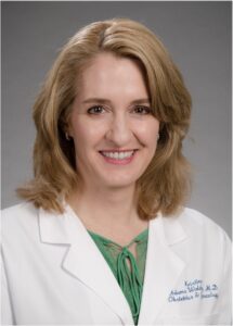
WaNPRC’s Interim Assoc. Director for Research, Kristina Adams Waldorf collaborated on a four-part series with other researchers in The Lancet Infectious Diseases and The Lancet Microbe. The series identified key research priorities needed to detect and mitigate the threat of future mosquito-borne Zika virus outbreaks. 

Dr Adams Waldorf is a leading researcher on Zika virus, which emerged in the Americas in 2015 and resulted in a devastating epidemic of infants born with small heads (microcephaly) and other severe congenital malformations. 

The four manuscripts focus on: Zika research priorities for preparedness and response, vaccines and monoclonal antibodies, non-human primate models of Zika virus, and sharing of specimens and data to accelerate Zika research and development. 

In the paper in which she was the senior author, the role of NHP models in research and developing zika countermeasures, she notes that Zika virus remains a threat to global pregnancies, is now endemic in 92 countries, and can be found in mosquitos in another 60 countries. 

Dr Adams Waldorf writes that developing therapeutics against Zika requires nonhuman primate research to mirror the physiology of human pregnancies.  NHP pregnancy is “remarkably similar to human pregnancy” she writes, from the interface between the fetus and mother to the fetal development. 

NHPs have emerged as the gold standard model for understanding the pathogenesis of ZIKV infection in humans and human pregnancy. Accelerating research and discovery on ZIKV will continue to rely on the availability of diverse non-pregnant and pregnant NHP models that can address different aspects of viral pathophysiology,” she writes. 

Another paper in the series notes that there are no licensed Zika vaccines or monoclonal antibodies currently available, which means world’s populations, particularly those who may become pregnant, are unprotected from Zika transmission, infection, and disease. 

Dr Adams-Waldorf’s paper can be found here: Role of non-human primate models in accelerating research and developing countermeasures against Zika virus infection published in The Lancet Microbe.  https://www.thelancet.com/journals/lanmic/article/PIIS2666-5247(24)00298-2/fulltext 

The other papers are: 

Zika virus vaccines and monoclonal antibodies: a priority agenda for research and development: https://www.thelancet.com/journals/laninf/article/PIIS1473-3099(24)00750-3/fulltext 

Zika virus: advancing a priority research agenda for preparedness and response:  https://www.thelancet.com/journals/laninf/article/PIIS1473-3099(24)00794-1/fulltext 

Specimen and data sharing to advance research and development on Zika virus:  https://www.thelancet.com/journals/lanmic/article/PIIS2666-5247(24)00325-2/fulltext 

Why I Work in Animal Research

This Guest Director’s Blog is by Mark Walton, PhD.
He is a senior research scientist at WaNPRC in the Neuroscience core. We welcomed his timely thoughts in a climate in which it’s valuable to understand the importance of animal research in advancing human and animal health.


In January of 2002, my first child, a son, was born. When he was still only minutes old, the doctor handed me our newborn child and said, “Here’s your son.” The joy I felt in that moment was beyond description, and I couldn’t help speculating about what the future would hold. What sort of life would he lead? Would I be up to the task of preparing this utterly helpless person for the independence of adulthood? I desperately hoped that he would one day find a career path that he would find fulfilling. I thought, too, about the fact that this newly arrived human being that had been entrusted to our care would likely outlive us by at least a couple of decades. I hoped that he would one day find love so that he would always have a family that he could trust.

As I held him in my arms for the first time, I could see that his skin had a bluish tint, but I wasn’t worried at first. We had been warned that newborn babies often take a little while to get their natural color, as their delicate little lungs begin to supply oxygen for the first time. I was so engrossed in the tiny miracle that rested in my arms that I failed to notice the growing concern that must have shown on the faces of the medical professionals that surrounded me. My own concern began to grow slowly, an empty feeling in my gut as I waited for the bluish tint to his skin to turn pink. Instead, his color worsened until a nurse gently took him from me. Soon a doctor explained to me that our son was having trouble breathing because his lungs were not as mature as they should have been. My wife and I were told that he had been taken to the neonatal intensive care unit (NICU), where they would help him to breathe. The technical term for this is respiratory distress syndrome.

That night my wife and I somehow managed to get to sleep. In the morning, the doctor told us that our baby had a “rough night.” She explained that normal lungs produce something called surfactant, which essentially keeps the lungs from sticking together when we exhale. To understand the need for surfactant she asked us to imagine trying to inflate a balloon that was wet on the inside. Our baby’s lungs, she said, were not mature enough to produce the surfactant that he needed. During the night, when he tried to inhale, one of his lungs stuck together. This ripped a hole in the lung, which caused it to collapse.

I listened, doing my best to show a strength that I didn’t feel. I knew this would be even worse for my wife, who was listening to the same horror story after giving birth to a baby. After the doctor finished, I stepped out into the hallway. The moment the door closed the fear that I had so carefully hidden came pouring out like flood water from a collapsed dam. In the hallway of the maternity ward – which should have been filled with joy – I sank to the floor, unable to stand. I wondered if our son was going to die without being held by his mother even once. I must have been quite a sight: a six-foot-three man crying uncontrollably on the floor while new parents walked past me with their healthy babies.

Later that morning, my wife and I were told that we could go to see our son. The doctor led us to the NICU but paused before opening the door. “I need to tell you what you are going to see,” she said. “Your son has a little ‘boo-boo’.” She used those words to describe what turned out to be a hole in our baby’s chest, into which a tube had been inserted. I remember watching the little drops of our child’s blood moving back and forth inside the semi-transparent tubing.

I couldn’t help wondering what our tiny son, just one day old, thought of the world that he’d entered. To him, it must surely be a place of endless pain and suffering. He hadn’t known comfort or love, except for those few precious minutes, in my arms. I would have done anything – even volunteered to be tortured to death – to take away his suffering and heal him.

In the days that followed, our son received an artificial surfactant, developed through years of research. It worked like a miracle. Soon, we were able to bring him home. That was 2002. Today he is a college student with a brilliant mind, studying astrophysics. He got married last summer, and I can honestly say that I have never seen a happier couple.

This miracle happened because of decades of animal research. Thanks to this research, scientists and medical doctors know a lot about how lungs work. Some of the most important work was done by studying pig lungs – experiments that could not have been performed on humans. Thanks to medical research involving animals, scientists learned the crucial role that surfactants play, and they learned how to make the artificial version that saved our son’s life. Before trying it on human babies, scientists first tested it on prematurely born monkeys. All medical treatments undergo this critical phase of testing, ensuring that new treatments are safe before they are given to humans – or, more personally, our children.

In 1963, President John F Kennedy’s newborn son tragically died of the very same problem that nearly killed our son. At that time, medical science had not advanced enough to save him. Medical research, including research involving monkeys, is the only reason that our son survived. For us, this is not a political issue; it is the difference between life and death. Yet as I write this, countless other families are not so fortunate. At this very moment, thousands of parents are watching helplessly as their children suffer and die of diseases that we have yet to treat. For them, the horror that I felt for a few short days will become an emotional wound that never heals.

It’s normal in a democracy for well-meaning, patriotic people to disagree about many things. The need to spare our fellow human beings the indescribable horror of losing a loved one should not be one of them. If there is one thing that we all should agree on, it is that we must support medical research. Every single person reading these words should remember that one day it might be your life that hangs in the balance, or the life of someone that you love. It is my great hope that we continue to support medical research so that an effective treatment will be found before that day comes for your family.

Orsborn Paper: Neural Information Could Impact Brain-Computer Interfaces

Photo of a smiling Amy Orsborn on a purple and gold background.
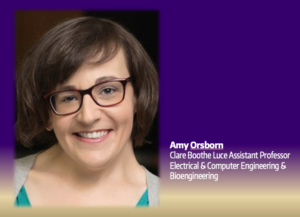
Neuroscience core scientist Amy Orsborn published a new paper in the high-impact publication Nature Neuroscience in which she, as lead author, reports that, “Neural populations are dynamic but constrained,” as the title reads. 

“Our brains evolved to help us rapidly learn new things. But anyone who has put in hours of practice to perfect their tennis serve, only to reach a plateau, can attest that our brains aren’t infinitely flexible,” the paper begins.  “New work shows that patterns of neural activity over time — the temporal dynamics of neural populations — cannot change rapidly, suggesting that neural activity dynamics may both reflect and constrain how the brain performs computations.” 

The findings, Dr Orsborn says, has the potential to impact how brain-computer interfaces are developed that could help provide people the use of artificial limbs. 

In the study, Orsborn and her colleagues used micro- electrocorticography recordings in two male monkeys to map how their eye movements related to their arm movements. The upshot is the movements mapped in the brain across different regions, which advances our understanding of how our brain works when we do everyday tasks like reaching toward things we’re not looking directly at. 

“These insights…reveal opportunities to leverage these signals to enhance future brain-computer interfaces,” she writes. 

Dr Orsborn is one of three University of Washington faculty members who recently received fellowships from the Alfred P. Sloan Foundation. Sloan Fellowships honor early-career researchers whose achievements mark them among the next generation of scientific leaders. Each fellow will receive $75,000 to apply toward research endeavors.

WaNPRC Highlights at NHP AIDS Conference

Logo for the Tulane National Primate Research Center Annual Symposium on Nonhuman Pimate Models for AIDS. It shows a trolly car with a red ribbon logo on the front

The 41st Annual symposium on Nonhuman Primate Models for AIDS is under way in New Orleans this week, and the Washington National Primate Research center is well-represented by both speakers and attendees.

The WaNPRC contingent consists of session speakers, poster presenters and attendees.  “This meeting is the only one of its kind in the world,” said Dr. Kristina Adams Waldorf, Interim Director of Research at WaNPRC.  “The symposium includes research presentations on HIV, HIV-like viruses, such as SARS-CoV-2, influenza, Zika, Valley Fever and others that nonhuman primates serve as a close model to human infection that can be used in nonhuman primates to model AIDS,  With 38 million people currently estimated to be living with HIV worldwide, the need to develop better treatments and interventions to prevent and/or cure HIV infection remains critical.”

Dr. Chris Peterson, a WaNPRC Affiliate with the Fred Hutch Cancer Center, is presenting on research targeting an HIV-like virus in macaques with (chimeric antigen receptor) CAR-T cells that are specific for a viral protein. “A major challenge to cure HIV is that antiretroviral therapy suppresses the virus to the point that it is essentially invisible to the immune system, but is not curative,” Dr. Peterson said. “What we’re trying to do here is extend how long our CAR T-cells work in the body, so that they more time to locate these rare, infected target cells, leading to a cure.”

Also presenting is Professor Donald Sodora, PhD, Adjunct Professor of Global Health, on the impacts of an HIV-like virus on the liver of nonhuman primates.

WaNPRC was important in establishing the symposium which began as a way for all seven NPRCs to meet and discuss research on the AIDS pandemic using nonhuman primates. At the time, primates were the only animal model that could be infected with a virus that was similar to HIV and develop AIDS symptoms.

“Back then, the NPRCs had been around for about 10-15 years, and were mostly focused on neurosciences,” said WaNPRC Director Dr. Deb Fuller. “But several scientists in infectious diseases at WaNPRC and others at Tulane first showed that old world monkeys (rhesus macaques at Tulane and pigtails at WaNPRC) could be infected with SIV. This launched a huge influx of funding from the NIH into the NPRCs to lead research in NHPs on AIDS and to this date, NIAID is still a major contributor to the P51 and U42 center grants that each NPRC is funded under to support their robust AIDS research programs.”

and has become a meeting place for a large number of scientists and Early-Stage Investigators, many of whom will give their first presentations.

One WaNPRC presenter is Orlando Cervantes, a graduate student in the Adams Waldorf Laboratory. He’ll be sharing his findings on how pandemic influenza infections during the third trimester of pregnancy damage the placenta by provoking a strong immune response.

“Scientists and doctors still don’t fully grasp the ways influenza infection can lead to stillbirth or preterm birth in pregnant women,” said Orlando. “By better understanding the placenta’s response to a maternal influenza infection, we can get clues on how that can compromise placental integrity and hurt the developing fetus. I’m very excited to share these findings because they can encourage other scientists to begin using NHP models to study the interaction between pregnancy and other illnesses/infections.”

The symposium follows on the heels of the HIV Vaccine Trials Network Early Stage Investigator conference, which also took place at the same location. The conference aimed at early-stage investigators (ESIs) specializing in translational HIV research in non-human primates (NHPs) and clinical HIV and TB research is set to offer participants valuable opportunities for professional growth. Attendees will gain insights into their potential contributions to the field and receive support in achieving key career milestones. Previous participants have reported that the event enhanced their skills and knowledge, while also providing a platform for building new collaborations.

Several WaNPRC researchers will have posters at the meeting including Director Deborah Fuller, Associate Director of Research Dr. Kristina Adams Waldorf, and Dr. Charlotte Hotchkiss, D.V.M, Ph.D. As well as the following:

  • Megan O’Connor – HIV/COVID-19 and/or HIV/ZIKV co-infection
  • Chris Peterson – HIV
  • Serena deBanco – HIV (scholarship recipient)
  • Megan Fredericks – Valley Fever

The symposium concludes October 25.