How mRNA and DNA vaccines could soon treat cancers, HIV, autoimmune disorders and genetic diseases
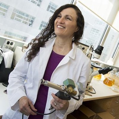
Fuller holding gene gun used to micro-inject DNA into skin cells
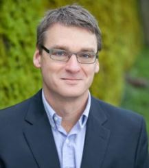
The two most successful coronavirus vaccines developed in the U.S. – the Pfizer and Moderna vaccines – are both mRNA vaccines. The idea of using genetic material to produce an immune response has opened up a world of research and potential medical uses far out of reach of traditional vaccines. Deborah Fuller is a microbiologist at the University of Washington and Associate Director of Research for the Washington National Primate Research Center. She has been studying genetic vaccines for more than 20 years. The Conversation spoke to her about the future of mRNA vaccines for The Conversation Weekly podcast.
Virus samples from Washington’s three positive omicron cases of COVID-19 are now in the hands of scientists in a super secure, Level 3 containment lab at UW Medicine’s complex of research centers in Seattle’s South Lake Union neighborhood.
“Do current antibodies from vaccination protect against omicron?” said Dr. Michael Gale about the upcoming experiments. Gale is the UW Medical School’s co-director for Emerging and Infectious Diseases, and Director for the Center for Innate Immunity and Immune Disease. His department also works with the National Institutes of Health. Continue reading…
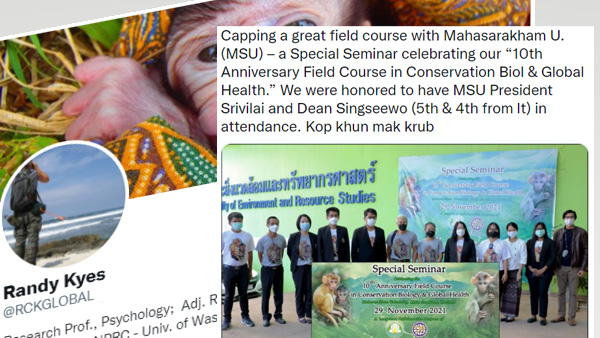
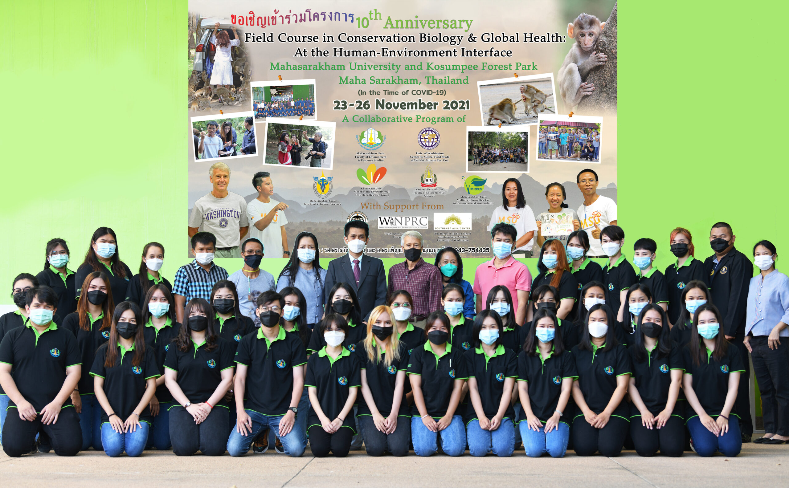
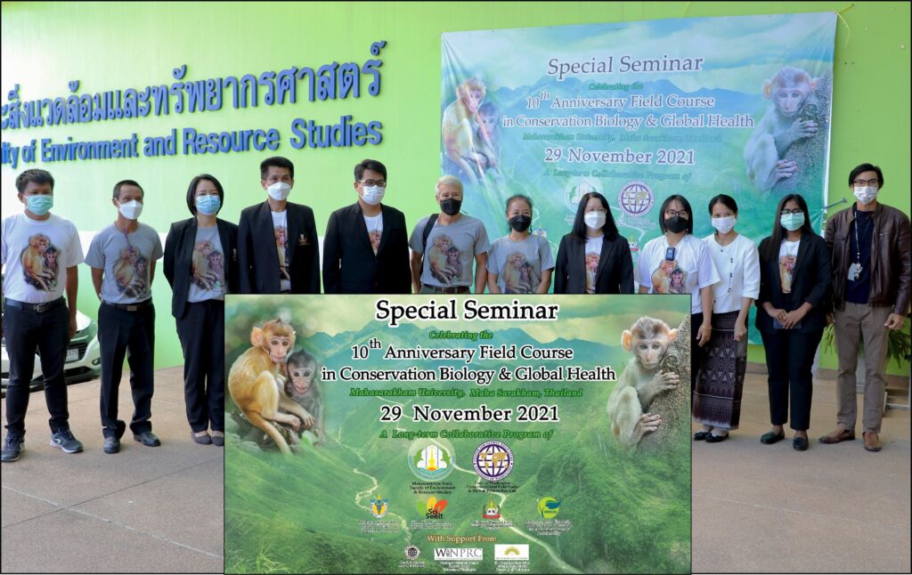
“We have managed to conduct this annual field course despite the pandemic – both last year and this year. And it was somewhat bittersweet, as this is the only field course (across all our program countries) that we will be able to conduct in 2021. Mahasarakham University also hosted a special seminar to celebrate the 10th anniversary of this field course. The past 20+ months have been a real challenge. Thank you to all the wonderful MSU students and staff.”
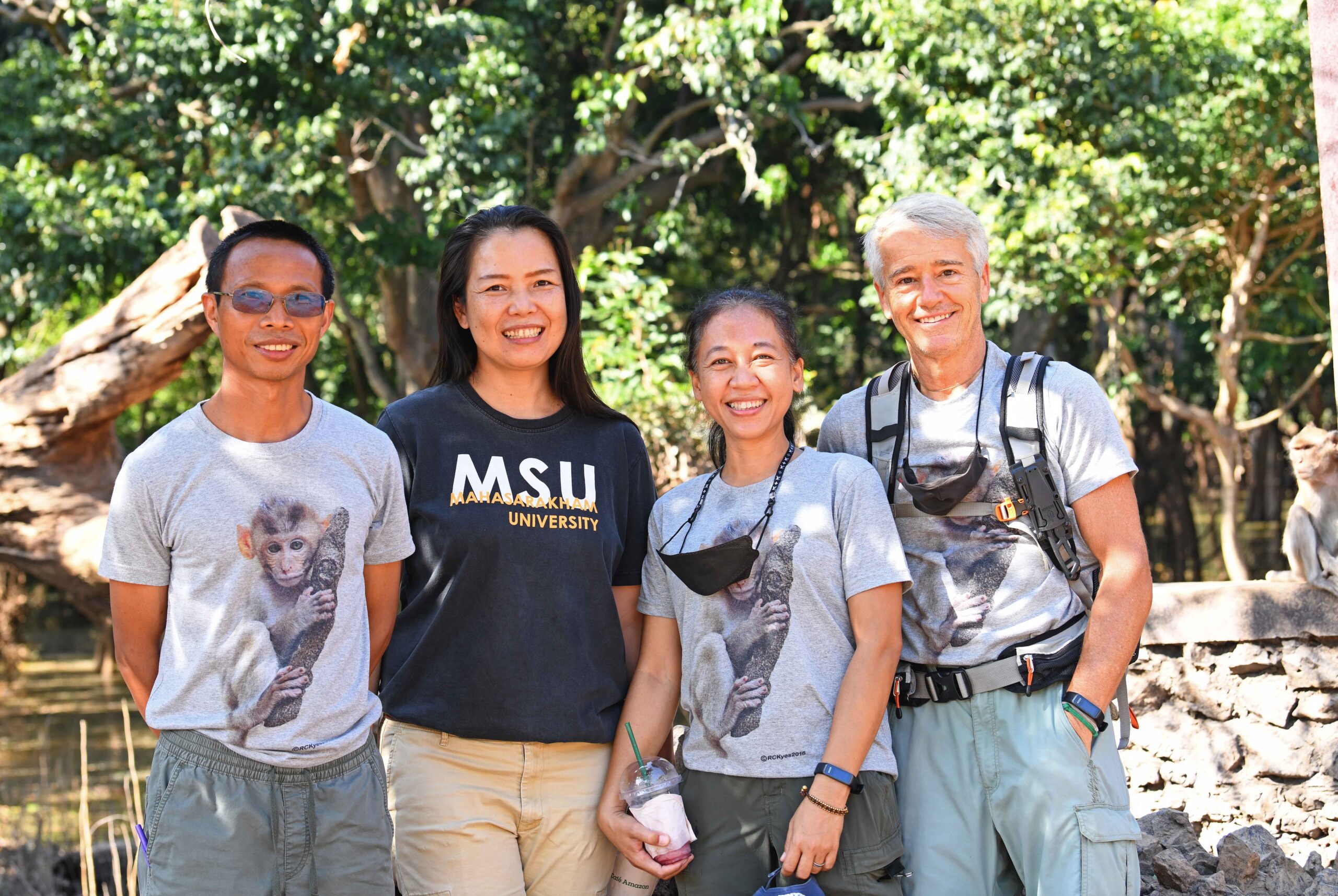
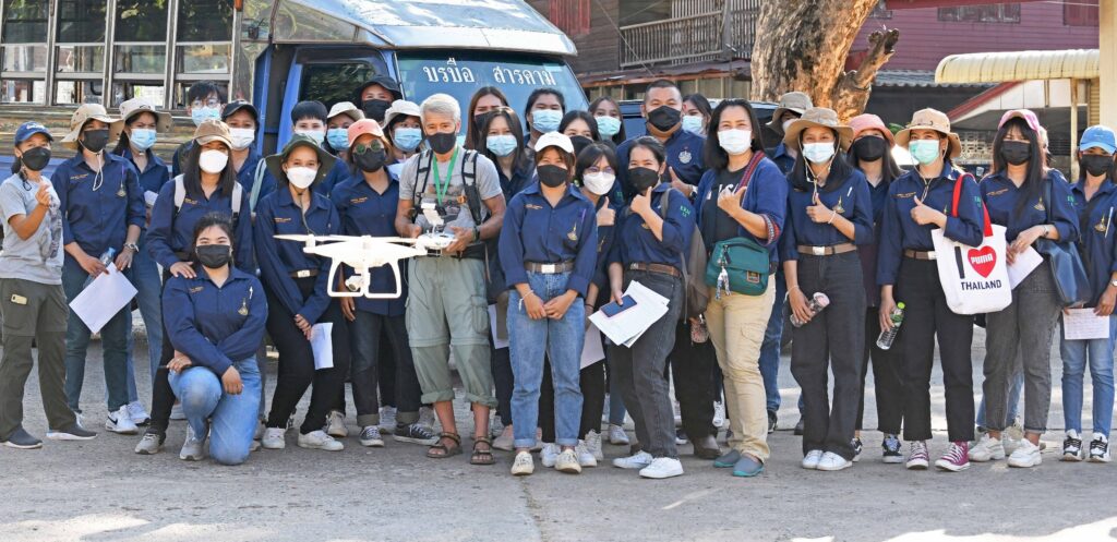
Great first day of our 10th anniversary Field Course in Conservation Biology & Global Health with our partners at Mahasarakham University, Thailand. The Team. It has been a true pleasure working with Dr. Tanee, Dr. Thamsenanupap, (Mahasarakham Univ., Thailand), and Dr. P. Kyes (WaNPRC @UW) – beginning with our very first field course in 2012. As we celebrate our 10th anniversary Field Course in Conservation Biology & Global Health, I want to express my gratitude to this team and all the others who have made this program a great success. Demonstration of an aerial drones and applications for conservation biology and global health. Special Seminar celebrating our “10th Anniversary Field Course in Conservation Biology & Global Health,” joined by Dean Singseewo (fourth from left) and MSU President Srivilai (fifth from left).
While the world’s newest coronavirus variant of concern, named omicron, hasn’t yet been detected in Washington state or the United States, a UW scientist said Friday that its high number of mutations is particularly concerning.
The new variant — which was first identified in South Africa and has now been seen in travelers to Belgium, Botswana, Hong Kong and Israel — has about 50 mutations, about 30 of which are located in the spike protein, a primary protein the virus uses to enter our cells, said Dr. Deborah Fuller, a microbiologist at UW Medicine.
“The concern regarding the number of mutations in that region is that there’s a potential that those mutations could make our vaccines less effective because the antibody response induced against the spike protein might be less effective against those mutants,” she said. Continue reading…
After a national search for a new center director, the WaNPRC has been awarded with the recruitment of an internationally recognized outside candidate. Effective October 16, 2021, Michele A. Basso, PhD, will join the WaNPRC as the new director.
Dr. Basso is already discussing plans for recruitment of new investigators and bringing in new core staff, particularly in the field of HIV and at the intersection of neuroscience and infectious disease.
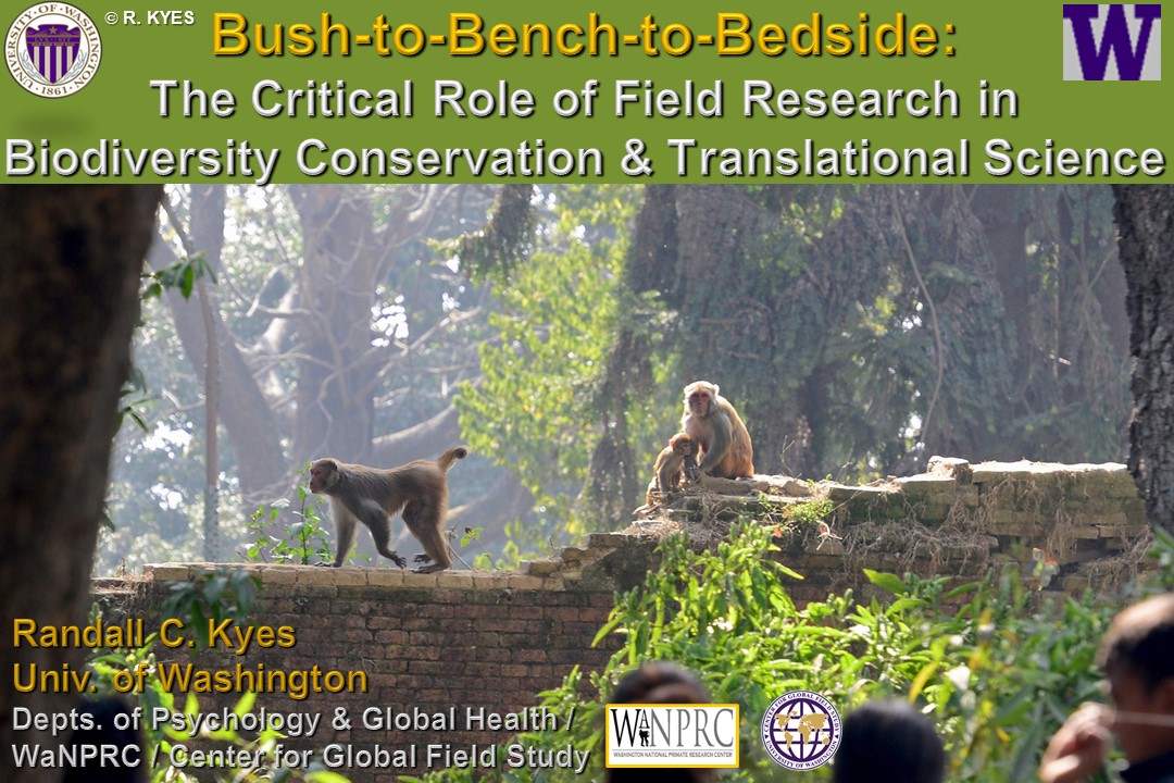
The Natural Resource Management Program at Nepal College of Engineering (NEC), in association with University of Washington, organized an August 20 webinar entitled, “Bush to Bench to Bedside: The Critical Role of Field Research in Biodiversity Conservation and Translational Science.”
The keynote speaker at the webinar, Professor Randall C. Kyes, said that long-term monitoring is very important to find out the status of biodiversity. He presented his research experience in different countries and emphasized on possible zoonotic diseases passing between humans and wildlife. Speaking about research on methicillin-resistant Staphylococcus aureus (MRSA) in monkeys of Kathmandu Valley. The Master of Sciences in Natural Resources Management program at the Center for Postgraduate Studies (CPS) is a collaborative partner for this research. Kyes said that evidence of human-to-human transmission of MRSA bacteria has been found in monkeys of Kathmandu Valley.
Warning of the potential dangers to wildlife from human and wildlife interactions, he said there is a need for ongoing research on wildlife and biodiversity. The webinar was attended by graduate students from various colleges. NEC CPS regularly organizes a series of webinars on various topics of biodiversity conservation and natural resource management.
The above is a translation of an article from Nagarik News, a Nepali language daily newspaper and news network, Nepal Republic Media Pvt. Ltd.
Dr. Randall C. Kyes is chief of the Division of Global Programs at the Washington National Primate Research Center.
As the global COVID-19 pandemic continues, safe and effective vaccines are playing a pivotal role in preventing severe disease and death and limiting the spread of SARS-CoV-2, the virus that causes COVID-19. The urgency of the COVID-19 pandemic necessitated rapid vaccine development and testing. Fortunately, NIAID’s decades-long support and conduct of coronavirus and vaccine research laid the groundwork for helping to develop a safe and effective COVID-19 vaccine in record speed.
COVID-19 Animal Models
Animal research plays a key role in developing successful vaccines for humans. Before promising vaccine candidates can be tested in humans, they must first be tested for safety and effectiveness in animals as required by the U.S. Food and Drug Administration. To do this, scientists first determine whether a vaccine candidate can stimulate an adequate and safe immune response. This important step is often conducted using small and then, potentially, larger animal models of disease. Mice are frequently used because they reproduce rapidly, have a well-characterized immune system and a defined genome. Some labs turned to mouse models of infection early in the COVID-19 pandemic only to find that mice don’t get infected with SARS-CoV-2. In order to infect cells, SARS-CoV-2 must bind to a human protein called ACE2. The human and mouse ACE2 proteins are different, and SARS-CoV-2 does not bind to mouse cells. Scientists overcame this problem by generating mice that can express the human version of ACE2 and can therefore be infected with SARS-CoV-2. When these genetically modified mice are infected by the virus, they lose weight and become sick in ways that are similar to what happens when people are infected with the virus. Mouse models provided vital information about COVID-19 symptoms and its disease course and continues to be used by researchers to understand COVID-19 disease.
Syrian hamsters are another important animal model for COVID-19 because disease in those animals closely resembles the disease in humans. Additionally, older male hamsters develop more severe disease than young female hamsters, which reflects some of the differences seen in humans infected by SARS-CoV-2. Hamster models have contributed to the evaluation of investigational COVID-19 vaccine candidates, immunotherapies, and antiviral drugs.
Vaccine development for COVID-19 also benefitted from nonhuman primate studies. In assessing immunogenicity and protection of vaccines in pre-clinical animal models, nonhuman primates provide several advantages for clinical translation. They are outbred, have greater similarity to humans than rodents in innate immune responses and B- and T-cell repertoires, and allow use of clinically-relevant vaccine doses. Recent studies in nonhuman primates show that SARS-CoV-2 targets similar replication sites and recapitulates some aspects of COVID-19 disease. Nonhuman primates are used during the later stages of vaccine development and typically build upon the knowledge accumulated in earlier small animal studies.
Professor Michael Manookin works at a confocal microscope.
Finding helps explain how baseball players can connect with a 100-mph fastball and how the rest of us manage everyday tasks.
Neural circuits in the primate retina can generate the information needed to predict the path of a moving object before visual signals even leave the eye, UW Medicine researchers demonstrate in a new paper.
“The ability to predict where moving objects will go is so important for survival that it’s likely hardwired into all sighted animals,” said Michael Manookin, an assistant professor of ophthalmology at the University of Washington School of Medicine. He led the research team with Fred Rieke, professor of physiology and biophysics.
Manookin and his colleagues report their findings in the journal Nature Neuroscience. Belle Liu and Arthur Hong, two UW undergraduate students, were the lead authors on the paper.
In the study, the researchers looked at how motion was processed by cellular circuits in the retina. The circuits the researchers focused on are composed of light-sensing photoreceptor cells, called cones; an intermediate layer of cells, called bipolar cells; and ganglion cells that collect signals from bipolar cells and transmit these signals out of the eye to other brain regions.
Ken Gordon, executive director of the Northwest Association of Biomedical Research (NWABR)
Just like our doctors and nurses, researchers in the Pacific Northwest have also been on the frontlines working at a rapid pace to understand the novel coronavirus that causes COVID-19: how it spreads, how it infects, who it infects, why it shows itself in a variety of symptoms and why it progresses in such a deadly way for some people.
Now more than 175 million Americans, and nearly 70% of eligible Oregonians, are directly benefiting from the biomedical research that developed these vaccines, bringing an end in sight to the pandemic.
Sadly, some animal rights organizations have used misinformation and fake news to disparage the biomedical research process during the past year.
Before anyone can be treated or vaccinated in America, medicines go through a lengthy process of lab research, humane animal modeling and human studies to ensure they work and are safe.
For the coronavirus vaccines this work started in libraries as researchers scoured the literature on coronaviruses and looked at decades of prior animal and human studies on vaccines. With this information in hand, researchers moved to their laboratories, where they identified ways to stimulate immunity to COVID-19.
The next step involved humane animal modeling to see if lab-developed products worked in living organisms. These animal studies typically started with mice, and if the results looked good then they proceed to working with animals like monkeys, which share more similarities with humans.
The last step involved human studies that started initially with just a few people and moved to trials that involved tens of thousands of volunteers. These studies were aimed at determining both the safety and the effectiveness of these vaccines.
We are all incredibly lucky researchers were able to develop three (with more on the way) highly effective COVID-19 vaccines in such a short time. Vaccine development work is still progressing with researchers at the Oregon Health and Science University working on the next generation of vaccines for COVID-19.
I understand how all this work created an existential crisis for animal rights groups, who oppose the use of any animals in research. But that’s no excuse to spin twisted stories about research and intimidate researchers.
Millions of Americans have directly benefited from ethical biomedical research and are now protected from severe symptoms and hospitalization due to COVID-19. Without animal studies we would not have these vaccines and we would still be in the thick of one of the worst pandemics in history.
Even worse, an animal rights supporter at a local university recently said he would prefer to have millions of people die if it meant an end to animal studies. Another anti-research activist compared animal researchers to child traffickers.
The people who are suffering the most from COVID-19 are people of color, people with underlying health conditions, people who are older and people with disabilities. These are people that seemingly some anti-research activists would throw under the bus.
I encourage people to be critical thinkers. Do your research and check facts. We live in a democracy where we can robustly debate issues. However, robust debate does not give activists the right to make up alternative facts.
Every academic research university, hospital and non-profit research institution in the Pacific Northwest has joined this fight against COVID-19. Just like the doctors and nurses on the frontlines, the hard work and dedication of these biomedical researchers saves lives and ensures we have a healthier community.
All 175 million of us who are vaccinated in America, and the many who are still protected even though they cannot get vaccinated due to underlying health conditions, should say #ThanksResearch to our local universities and research institutions who are saving lives.
Don’t buy into lies from those who bitterly reject science.
Ken Gordon is executive director of the Northwest Association of Biomedical Research.
This opinion piece also appeared in the Portland Tribuneon July 02, 2021
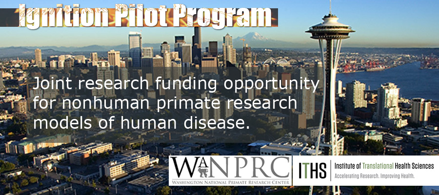
The WaNPRC Pilot Program is conducted jointly with the Institute for Translational Health Sciences and provides funding to collect preliminary data for future funding opportunities. The goal is to fund projects with innovative research endeavors that have translational implications to move toward human applications. This year there was a competitive pool of applications and we were able to provide funding to 2 projects.
Please join us in congratulating the Grant Year 60 recipients of the WaNPRC Pilot Program which exemplify the commitment to cutting edge science, collaboration and also support the 3Rs (Reduction, Refinement and Replacement) of animal use.
Jesse Erasmus, PhD, (University of Washington, Fuller Lab) “Overcoming bottlenecks in mRNA-mediated antibody expression in nonhuman primates”
Abstract: We have demonstrated protection from acute virus infection in mice receiving an intramuscular injection of RNA encoding a monoclonal antibody. While this approach holds promise for enabling rapid development of antibody therapeutics that can be administered in an outpatient setting, scaling intramuscular doses from mice to larger animals has proven difficult, failing in a nonhuman primate pilot study. We have since identified three major bottlenecks in antibody expression in vivo, 1) host translation shutdown mediated by endoplasmic reticulum (ER) stress and plasmacytoid dendritic (pDC) cell interferon production, 2) anti-drug antibody response, and 3) limiting numbers of transfected target cells. We have developed an RNA molecule that co-expresses a modified antibody along with a cell-autonomous human ICAM-1 blocking peptide, designed to reduce ER stress and inhibit local pDC interaction with RNA-transfected cells, respectively. Given the 87% similarity between human and pigtail macaque (PTM) ICAM-1, we hypothesize that in PTMs, this will 1) reduce ER stress- and pDC-mediated interferon production and downstream host translation shutoff, and 2) reduce pDC-mediated anti-drug antibody responses. In order to increase the number of target cells, we propose to use an FDA-approved multi-needle array in order to spread an injection volume over multiple sites.
Amy Orsborn, PhD, (UW Department of Electrical & Computer Engineering and UW Department of Bioengineering, WaNPRC Core Staff): “Developing and validating a new behavioral assay to quantify feedforward and feedback”
Abstract: Our movements are controlled by a combination of predictive feedforward control and reactive feedback control. Incorporating insights from feedforward/feedback control has significantly improved therapies to restore motor function like brain-machine interfaces. However, existing methods to study motor behaviors in non-human primates (NHPs) cannot quantify and disentangle feedforward and feedback control components. This methodological gap limits our ability to study the neural mechanisms of sensorimotor control. Robust control theory methods have been used to directly quantify feedforward and feedback sensorimotor pathways in humans, but these approaches have not yet been tested in NHPs. We propose a study to develop and validate these behavioral assays in NHPs. We will develop assays for both in-cage assays that may accelerate training (aim 1) and laboratory assays for more complex, higher dimensional movements (aim 2). With expertise in both NHP behavioral training and control theory, our team is positioned to rapidly generate critical feasibility data for future grants. If successful, our proposal will provide primate neuroscientists with new tools to study the neural mechanisms of sensorimotor learning and control. Quantifying feedforward and feedback control and their neural signatures will also enable improved brain-machine interface therapies.
We wish the recipients luck in their endeavors and we look forward to hearing about their exciting results next year.
Elizabeth A. Buffalo, PhD | Interim Associate Director for Research
The WaNPRC performs critical biomedical research leading to new advances in science and medicine. WaNPRC researchers are working to develop effective vaccines and therapies for HIV/AIDS and other infectious diseases as well as new advances in genetics, neuroscience, vision, and stem cell biology and therapy. The WaNPRC directly supports the National Institutes of Health’s mission to translate scientific advances into meaningful improvement in healthcare and medicine.