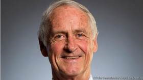
When and how did you first arrive here at the UW and Primate Center? What was your path to the position you are now leaving?
I graduated as a Vet Tech (Pierce College) in 1979. I was working at the old Emergency Vet Hospital with Bill Morton, who was also the WaNPRC Supervisory Vet at the time. He ‘recruited’ me into the WaNPRC, as the overnight, graveyard vet tech, in 1982. So my first few years here were passing out meds, treating minor injuries, checking lixits and water filters, assisting feeding times at the Infant Lab, and “other duties as needed.”
After I moved to daytime working hours, I had the opportunity to work in many areas of the Primate Center – daily clinical support, tissue program, surgery, etc. After being the Vet Tech assigned to the Infant Lab for some years in the late 80s/early 90s, I was offered the opportunity to work with the AIDS-related research projects. This was early in the AIDS outbreak and it was the policy to limit the personnel with access to the animals assigned to those protocols. So a small number of staff were responsible for the conduct of the experiments, data collection and the daily oversight of their clinical status. This was before the existence of the Research Support Services – actually, this small group became the ‘seed’ that the RSS sprouted from. It became clear that having consistent research support from staff that were comfortable with the animals, liked the animals and were a familiar presence to the animals, was a benefit to both the study and to the animals’ well-being.
The outgrowth of the research work developed into helping PIs develop the budgets for their studies. After a time, the hands-on animal work had to become a part of my past. The budget developments became a full-time effort assisting PIs with their pre-award processes, for scientists within the Center, other UW departments, and other institutions.
Looking back on your years at the Center, what are some positive changes you have seen in NHP research? What possibly has stayed the same?
I think the thing that really stands out to me is the recognition of the need for the Psychological Well-Being (PWB) and the Environment Enrichment programs. There had always been efforts made on the part of individuals. Having a mandated program, with coordinated supervision, has been a pleasure to observe. Even though I personally have not had any interactions with the animals in a number of years, I know the staff in the program are passionate about their work and the PWB of the animals is in good hands.
The hope is that we are constantly making things better, faster, smarter or less expensive. We try to strive to do more—with less. Perhaps tell us about a project or problem that you improved in these ways.
I’d like to think that I am a solution oriented type of person, with a eye out for areas that could benefit from changes (and coincidentally making my job easier). Some of these improvements are still being used today, albeit with updates and modifications as warranted. After all, every process, every form, every SOP should always be seen as a living thing with changes made as time, circumstances, rules and guidelines and technology move forward. There is no improvement that can’t be improved. I am proud that some of the seeds I have planted have shown value and have grown.
What’s next for you, Ann?
Disneyland?
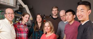
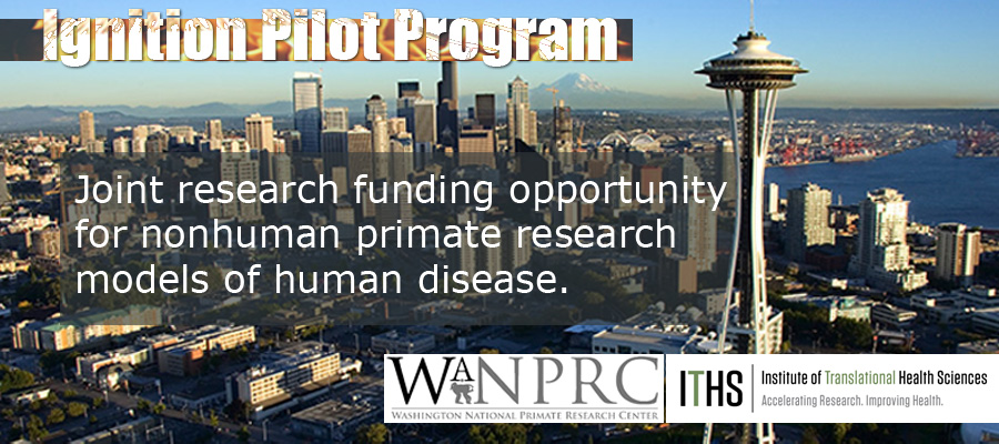 The WaNPRC Pilot Program, which is conducted jointly with the Institute for Translational Health Sciences, has been presented previously in an earlier Weekly Update. Since that time, the awards for the Fiscal Year 56 have been made and we want to present some short summaries of these protocols, to demonstrate the type(s) of research the Pilot Program supports. In short, the intent of the program is to support: